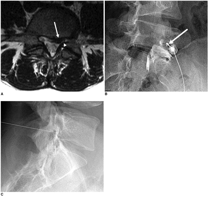
Fig. 2.
A 20-year-old girl with left leg pain. On the T2-weighted MR images (A), an extruded disc (arrow) was evident at L5-S1. This disc was located in the left central zone and it had migrated inferiorly to compress the left S1 nerve root (arrowhead). We performed transforaminal epidural injection with using the preganglionic approach at the L5-S1 level (B, C). In the oblique view (B), the needle tip was inserted just lateral to the pars interarticularis (arrow). The leg pain had been relieved at the 2-week follow-up.

