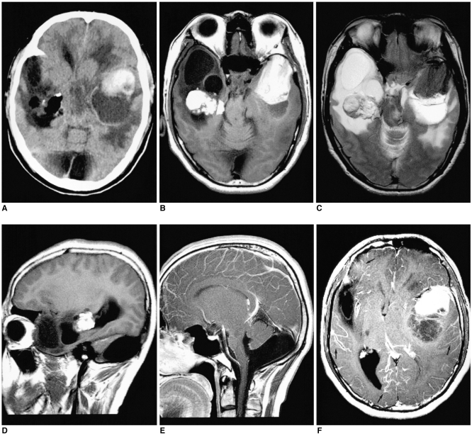
Fig. 1.
Neurocutaneous melanosis in a 27-year-old man.
A. Noncontrast CT scan demonstrates a hyperdense mass with an adjacent cyst in the left temporal lobe. The CT scan also shows an irregular fatty mass (-105 HU) with marginal calcifications within the temporal horn of the right lateral ventricle.
B, C. The axial T1-weighted (B) and T2-weighted (C) MR images show a left temporal lobe mass that is hyperintense on T1-weighted images and it is hypointense on T2-weighted images. There is a peritumoral cyst posterior to the main mass. The MR images also showed a mass in the right lateral ventricle, which appears homogeneously hyperintense on the T1-weighted images and heterogeneously hyperintense on the T2-weighted images; this is consistent with a dermoid cyst. The cystic encephalomalacia in the right temporal lobe is probably related to an early childhood insult.
D. The right parasagittal T1-weighted MR image confirms the location of the right side mass within the temporal horn of the lateral ventricle.
E. The midline sagittal contrast-enhanced T1-weighted MR image reveals hypoplasia of the inferior vermis and dilatation of the inferior fourth ventricle that communicates to the enlarged posterior fossa.
F. The axial contrast-enhanced T1-weighted MR image shows mild enhancements of the wall of the peritumoral cyst in the left temporal lobe. Also noted is mild diffuse enhancement of the leptomeninges (arrows).

