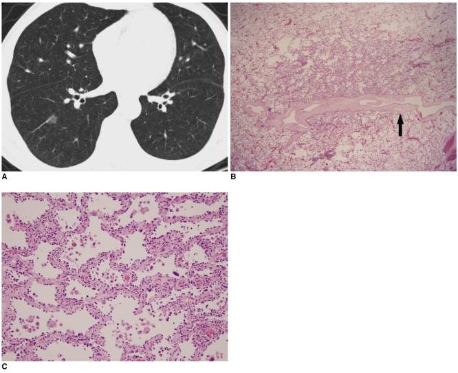
Fig. 2.
A 53-year-old woman with a single atypical adenomatous hyperplasia in the right lower lobe.
A. Transverse thin section CT scan shows an 11.2×10 mm well defined round nodule with pure ground-glass opacity in the right lower lobe. Note the pulmonary vessel penetrates the ground-glass opacity lesion without any vascular compromise.
B. Photomicrograph (H & E staining, ×10) shows that the boundary between the atypical adenomatous hyperplasia and the underlying lung parenchyma is distinct. Note the pulmonary vessel penetrating the AAH lesion (black arrow).
C. High power photomicrograph (H & E staining, ×100) shows atypical epithelial cell proliferation along the thickened alveolar septa, which is consistent with atypical adenomatous hyperplasia.

