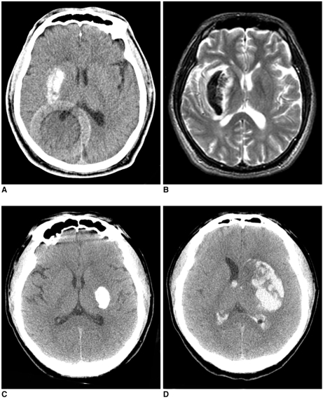
Fig. 5.
A 58-year-old man (A, B) and a 67-year-old man (C, D) show typical 'metallic high density' lesions with different outcomes.
A. The non-contrast CT obtained immediately after intra-arterial thrombolysis in the patient whose initial NIHSS score was eight. There is a very hyperdense lesion in the right basal ganglia with a CT value of 90 HU.
B. The follow-up gradient-echo MR image obtained two days later shows a hematoma without significant mass effect.
C. The non-contrast CT obtained immediately after intra-arterial thrombolysis in the patient whose NIHSS score was 17. A round lesion with very high density is noted in the left basal ganglia. The CT value was calculated as 260 HU.
D. The follow-up CT obtained after 24 hours shows a significant hemorrhagic transformation of the hyperdense lesion.

