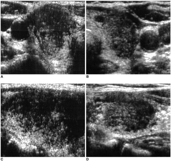
Fig. 1.
A 62-year-old woman with diffuse neck swelling and malaise. The laboratory tests suggested normal thyroid function. The transverse right (A) and left (B), and longitudinal right (C) and left (D) thyroid sonograms show ill-defined hypoechoic lesions involving nearly the entire area of both thyroid glands. Both thyroids are diffusely enlarged, but no cervical lymphadenopathy was detected. Subacute granulomatous thyroiditis was confirmed by fine needle aspiration biopsy. The patient's condition improved dramatically following steroid treatment.

