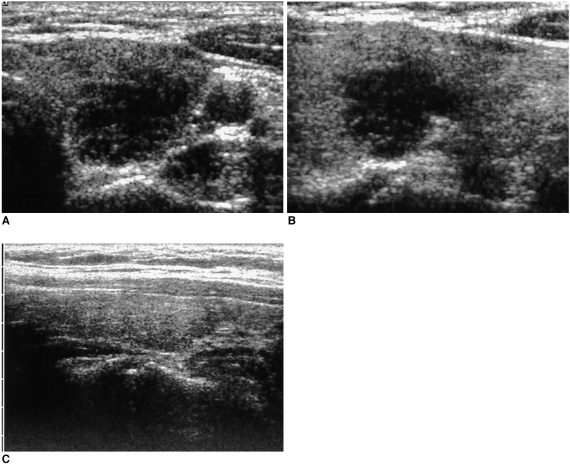
Fig. 3.
A 50-year-old woman with neck swelling. Transverse (A) and longitudinal (B) sonograms of the left thyroid show an ill-defined, markedly hypoechoic lesion mimicking a malignant nodule. Subacute granulomatous thyroiditis was confirmed by performing fine needle aspiration biopsy. On the follow-up longitudinal sonogram after one month of medication (C), the lesion is not clearly visualized.

