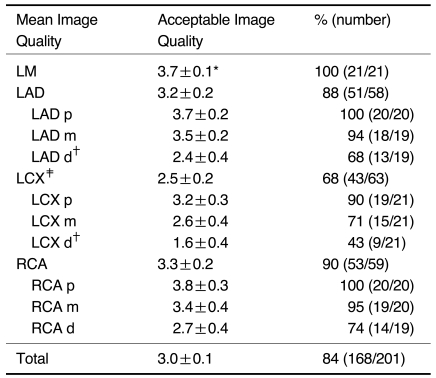
Table 1.
Quality of the Images of Whole Heart Coronary MRA
Note.-Image quality was graded as 0 = uninterpretable, 1 = poor, 2 = fair, 3 = good or 4 = excellent; *= standard error of the mean; †= Image quality of the LCX was significantly lower (p < 0.05) than that of the other arteries; ‡= Image quality of the distal segments of the LAD and LCX was significantly lower (p < 0.05) than that of the other segments; p = proximal; m = middle; d = distal

