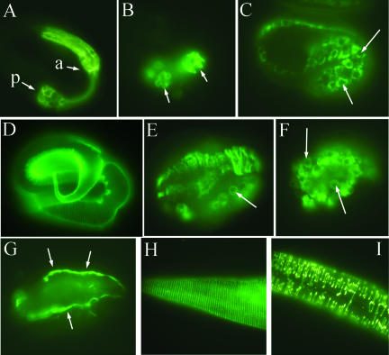
Figure 4.
Effect of sec-23 mutation on pharyngeal development and on the secretion of collagen and a basement membrane marker. (A and B) A pretzel-stage wild-type embryo (A) and a sec-23(ij13) mutant (B) were stained with antibody 3NB12, which labels pharyngeal muscle cells (Priess and Thomson, 1987). The anterior (a) and posterior (p) pharyngeal bulbs are indicated in A; the two bulbs are also indicated in B, but due to the severity of the morphological defects, it is not possible to distinguish anterior and posterior. (C–F) Immunolocalization of the DPY-7 collagen with the DPY7–5a mAb. (C) A wild-type late comma-stage embryo showing perinuclear localization of DPY-7 in hypodermal cells before secretion. Two nuclei are indicated by white arrows. (D) A wild-type pretzel-stage embryo with DPY-7 in the cuticle. (E) A sec-23(ij13) mutant embryo at terminal zygotic phenotype, with some DPY-7 retained in a perinuclear location (arrow) and most in aberrant strands or clumps. (F) Perinuclear DPY-7 (arrows) in a sec-23(ij13) mutant embryo derived from a chromosomally sec-23(ij13) homozygous mutant mother transgenic for sec-23. (G) Nonperinuclear localization of the basement membrane component perlecan with MH3 antiserum in a mutant embryo like that in F. (H and I) Localization of DPY-7 to circumferential bands in the cuticle of a wild-type larva (H) and to discontinuous bands in a sec-23 RNAi larva.

