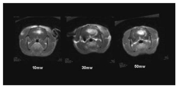
Fig. 5.

T1-weighted MRI contrast enhanced images showing focal contrast enhancement as evidence of BBB disruption. The effects of fluence rate on BBB disruption are clearly evident. Increased fluence rates resulted not only in increased contrast volume but increased signal intensity as well, indicating an increased contrast concentration.
