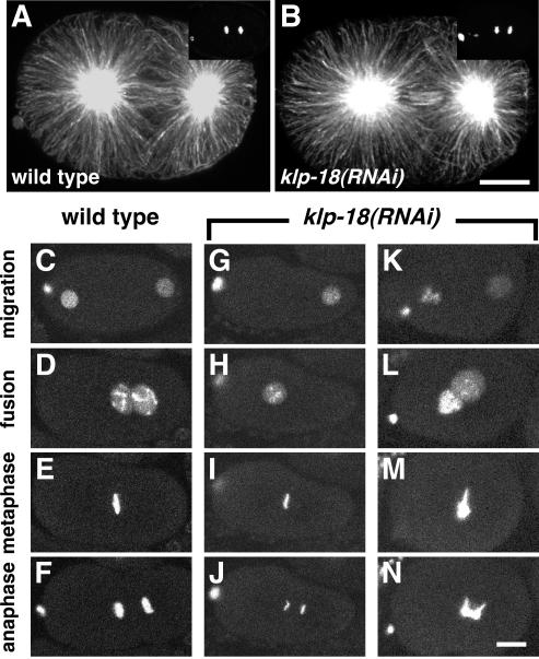
Figure 8.
Spindle organization and chromosome behavior during first mitosis in klp-18(RNAi) embryos. (A and B) Staining of the first mitotic spindle with α-tubulin antibodies reveals a similar pattern in wild-type and klp-18(RNAi) embryos. Yoyo-1 staining of DNA, shown in the top right corner, reveals that both embryos were in late anaphase. Anterior is to the left. (C–N) Time-lapse image series of GFP::histone H2B in live wild-type and klp-18(RNAi) embryos during pronuclear migration and first mitosis. (C–F) In wild-type, the female pronucleus migrates to meet the male pronucleus in the posterior hemisphere (D), the pronuclei move to the center of the embryo, and the chromosomes condense into a tight metaphase plate (E) and cleanly separate into two distinct masses during anaphase (F). (G–J) In this klp-18(RNAi) embryo an oocyte pronucleus did not form. The sperm-derived haploid complement of chromosomes underwent apparently normal mitosis (I and J). (K–N) In this klp-18(RNAi) embryo, the oocyte pronucleus contained an abnormally large amount of DNA. The oocyte-derived chromatin complement did not condense or congress normally at metaphase (M) and displayed bridges during anaphase (N). The sperm-derived chromosomes seemed to behave relatively normally. Bars, 10 μm.

