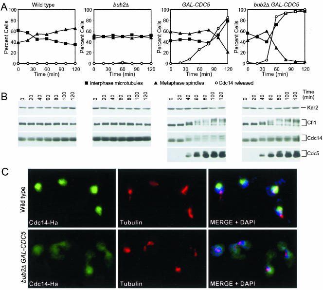
Figure 5.
Overexpression of CDC5 causes release of Cdc14 from the nucleolus in hydroxyurea-arrested cells, which is enhanced by the deletion of BUB2. Wild-type (A1681), bub2Δ (A4451), GAL-CDC5 (A4450), and GAL-CDC5 bub2Δ (A4453) cells carrying a CDC14-3HA and a CFI1-3MYC fusion were arrested in early S phase with HU (10 mg/ml) in YEPR medium at 25°C. After 2.5 h 5 mg/ml HU and galactose (2%) were added. Samples were taken at the indicated times to analyze the percentage of cells with metaphase spindles (closed triangles; A) interphase microtubules (closed squares; A), with Cdc14 released from the nucleolus (open circles; A) and to analyze Cfi1/Net1, Cdc14, and Cdc5 protein levels and their mobility (B). Kar2 was used as an internal loading control in Western blots. The pictures in (C) show Cdc14 localization in wild-type and GAL-CDC5 bub2Δ cells 80 min after galactose addition. Cdc14 is shown in green, microtubules in red and nucleus in blue.

