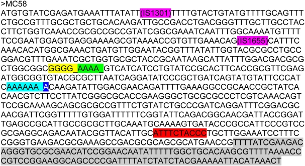
Figure 2. Presentation of all lpxL1 mutations.
The lpxL1 gene sequence of strain MC58 is shown including the different types of mutations and their positions in the gene found among isolates from patients. Each type of mutation is indicated with a different color. An ‘_’ indicates a nucleotide that is deleted in the mutant strain, pink indicates an insertion element (type I and II mutations), yellow indicates a deletion of a guanine in a stretch of five guanines (type III mutation), green indicates a deletion of an adenosine in a stretch of five adenosines (type IV mutation), light blue indicates a deletion of an adenosine in a stretch of seven adenosines (type V mutation), dark blue indicates an insertion of an adenosine in a stretch of seven adenosines (type V mutation), and the sequences that are highlighted in red or gray are deleted in the mutant strain (type VI and VII mutations respectively).

