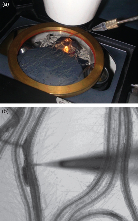
Figure 5. Experimental set-up for the microaspiration of infected Arabidopsis roots.
(a) A metal ring fixed under an inverse microscope (Zeiss, http://www.zeiss.com) holds a thin glass plate covered with medium enclosing the roots. (b) A microcapillary is navigated towards the roots by a micromanipulator (Eppendorf, http://www.eppendorf.com) for piercing a single syncytium.

