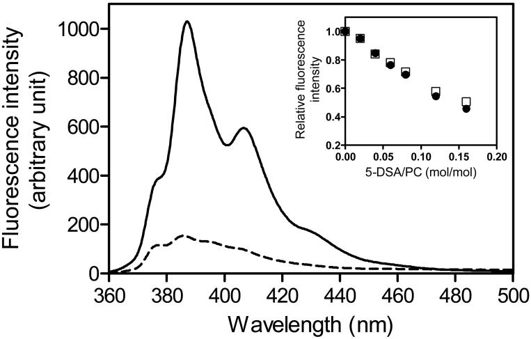
Fig. 2. Fluorescence emission spectra of pyrene-labeled apoA-I V53C mutant in the lipid-free (dashed line) or bound to egg PC SUV (solid line).
Protein and PC concentrations were 25 μg/ml and 1.0 mg/ml, respectively. The inset shows the relative changes in fluorescence of apoA-I V53C-pyrene (□) and F229C-pyrene (•) as a function of the molar ratio of 5-DSA to PC.

