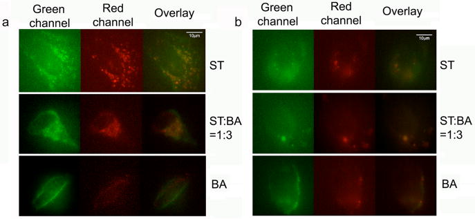
Figure 5.
Live-cell imaging uses fluorescent color-switching nanoparticles. (a) Adjusting the monomer ST-to-BA ratio effectively reduces non-specific interactions between nanoparticles and Hela cells. The residue green fluorescence in BA line is mostly due to cell autofluorescence. (b) Similar to (a) except that Hela cells are replaced with non-cancer HEK-293 cells. In both (a) and (b), nanoparticles only adhere to the cells when the monomer A and BA are out of balance, but undergo no endocytosis. The overlay images confirm the signals are from nanoparticles, not interference.

