Figure 1.
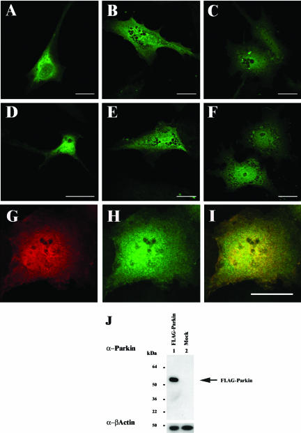
Localization of Parkin in neuronal and nonneuronal cell lines. FLAG-Parkin was transfected into SH-SY5Y (A and D), U138MG (B and E), or COS-7 (C and F–I). Forty-four hours post-transfection, the cells were fixed with 4% (wt/vol) paraformaldehyde, permeabilized, and immunostained with mouse monoclonal anti-FLAG (1:1000) (A–C and G), or affinity-purified rabbit anti-Parkin peptide (1:50) (D–F and H) antibodies. Anti-FLAG and anti-Parkin demonstrate overlapping staining patterns in COS-7 cells transfected with FLAG-Parkin (I, yellow staining indicates regions of colocalization within cells). Bars, 20 μm. (J) COS-7 cells lysates from FLAG-Parkin (lane 1) or mock (lane 2)-transfected cells were prepared in RIPA buffer 48 h post-transfection. Ten micrograms of each total cell lysate was separated by SDS-PAGE and analyzed by Western blotting with affinity-purified anti-Parkn C-terminal peptide (1: 500) and anti-β-actin (1:5000) antibodies. Arrow indicates the presence of FLAG-Parkin. A band corresponding to the expected position of endogenous Parkin was not observed in either lane.

