Figure 2.
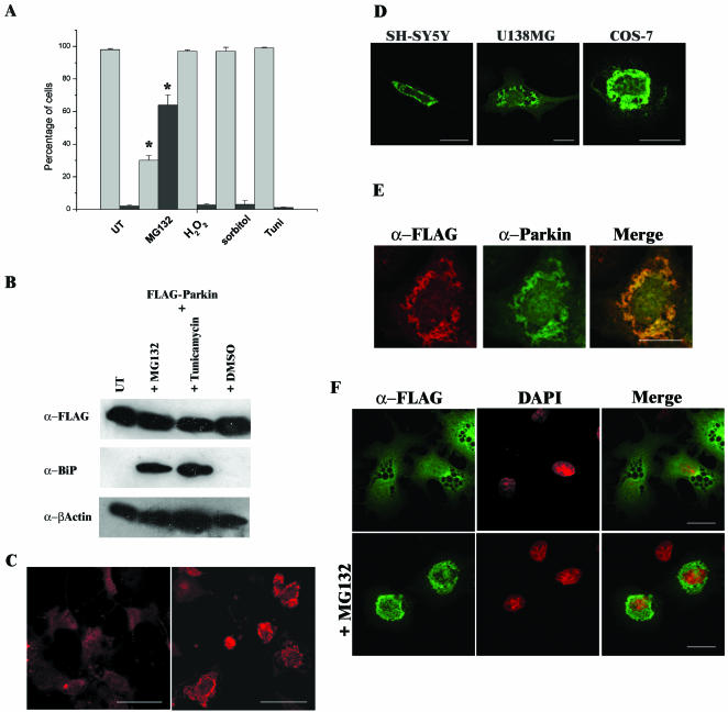
Addition of the proteasomal inhibitor MG132 causes inclusion body formation in cells overexpressing Parkin. (A) COS-7 cells were transfected with FLAG-Parkin. Twenty-eight hours post-transfection, cells were incubated for 16 h in the presence of either 20 μM MG132, 400 μM hydrogen peroxide (H2O2), 300 mM sorbitol, or 10 μg/ml tunicamycin. The presence of Parkin was then assessed by immunofluorescence with mouse monoclonal anti-FLAG antibody. Light gray bars represent the percentage of cells demonstrating a “wild-type” staining pattern, and dark gray bars represent the percentage of cells containing inclusion bodies. A minimum of 300 transfected cells was analyzed for each sample. Data shown represent the results of two independent sets of experiments. Error bars indicate the SE from the mean of these experiments. The asterisk (*) indicates a significant difference between the percentage of untreated cells versus the percentage of treated cells. *p < 0.01. (B) Tunicamycin and the proteasome inhibitor MG132 induce the UPR. COS-7 cell lysates from FLAG-Parkin–transfected cells left untreated (UT) or treated with 20 μM MG132, 10 μg/ml tunicamycin, or the carrier DMSO for 16 h were prepared in RIPA buffer 48 h post-transfection. Ten micrograms of each total cell lysate was separated by SDS-PAGE and analyzed by Western blotting with anti-FLAG, anti-BiP (1:250), and anti-β-actin antibodies. (C) COS-7 cells were transfected with FLAG-Parkin. Twenty-eight hours post-transfection, cells were grown in the presence or absence of 20 μM MG132 for 16 h before fixation. The presence of Parkin was assessed as described above. COS-7 cells containing MG132 induced inclusions (right) display a more intense staining pattern than untreated cells overexpressing Parkin (left). Confocal images of cells were scanned at the same fluorescent intensity. Bars, 50 μm. (D) Proteasome inhibition induces inclusion formation in SH-SY5Y, U138MG and COS-7 cells overexpressing FLAG-Parkin. (E) Immunostaining of inclusions with anti-FLAG (red) or anti-Parkin peptide (green) antibodies demonstrated similar staining patterns as indicated by yellow staining in merged panel. (F) Inclusions formed are within the cytosol. Transfected COS-7 cells were assessed by immunofluorescence by using the monoclonal anti-FLAG antibodies (green) and counterstained with 4,6-diamidino-2-phenylindole to visualize nuclei (red). Bars (D–F), 20 μm.

