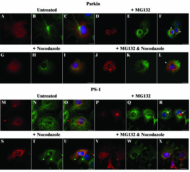
Figure 5.
Effect of nocodazole on Parkin and PS-1 inclusions. COS-7 cells were transfected with FLAG-Parkin (A–L) or PS1 (M–X) constructs and grown in the absence (A–C and M–O) or presence of 20 μM MG132 (D–F and P–R) or 10 μg/ml nocodazole (G–I and S–U) or both (J–L and V–X) for 16 h. After methanol fixation, cells were probed with affinity-purified rabbit anti-Parkin peptide (1:50) (A–L; red), or affinity-purified rabbit anti-PS1 peptide (1:50) (M–X; red) antibodies, and rat anti-α-tubulin (1:500) (A–X; green) antibodies. Overlays of each set of three include 4,6-diamidino-2-phenylindole staining (blue) to identify nuclei (C, F, I, L, O, R, U, and X). Regions of colocalization within cells stain yellow. Arrowheads indicate Parkin inclusions; arrows indicate PS1 aggresomes. Bars, 20 μm.

