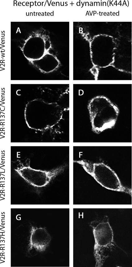
Figure 7.
Confocal microscopy using HEK293 cells that were untreated (A, C, E, and G) or treated with AVP for 30 min (B, D, F, and H). Cells expressed untagged β-arrestin 2 with V2R-wild type/Venus (A and B), V2R-R137C/Venus (C and D), V2R-R137L/Venus (E and F), or V2R-R137H/Venus (G and H) in the presence of dominant-negative dynamin, dynamin(K44A).

