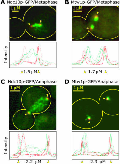
Figure 5.
Localization of Ndc10-GFP and Mtw1p-GFP in metaphase and anaphase cells. Ndc10p and Mtw1p, a known kinetochore protein (Goshima and Yanagida, 2000), were tagged with GFP (green) and the spindle pole component Spc42p was tagged with CFP (red; indicated by yellow arrowheads). Maximum intensity projections of three-dimensional image stacks containing 10 to 20 0.2-μm sections are shown representing typical images. The outline of the cell is indicated in yellow. All cells were exposed similarly and images have been adjusted to give the most accurate comparison. The graphs show the GFP and CFP fluorescence (raw pixel intensities) integrated along the spindle axis for three different representative cells with the solid line and indicated spindle length representing data from the image shown.

