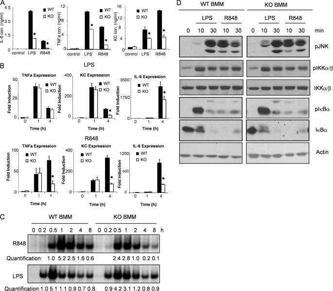
FIGURE 3.
TLR-mediated cytokine and chemokine production and NFκB activation in IRAK2-deficient macrophages. A, WT and IRAK2-deficient (KO) BM-derived macrophages were treated with LPS (1 μg/ml) or R848 (1 μg/ml) for 24 h. IL-6, KC, and TNF-α concentrations in supernatant were measured by enzyme-linked immunosorbent assay. The results shown are the means ± S.D. of triplicate determinations. *, p < 0.05. B, WT or IRAK2-deficient (KO) BM-derived macrophages were treated with LPS (1 μg/ml) and R848 (1 μg/ml) for indicated times, and total RNAs (2 μg) were analyzed by reverse transcription-PCR. *, p < 0.05. C, cell lysates from WT and IRAK2-deficient (KO) BM-derived macrophages, either untreated or treated with LPS (1 μg/ml) or R848 (1 μg/ml) for indicated times analyzed by electrophoresis mobility shift assay with an NFκB-specific probe. The levels of NFκB activation were analyzed by Scion Image 1.62C alias and are presented as fold increases compared with the samples treated for 30 min with R848 or 15 min with LPS. D, WT or IRAK2-deficient (KO) BM-derived macrophages were untreated or treated with LPS (1 μg/ml) or R848 (1 μg/ml). The cell lysates were analyzed by the Western method with antibodies against phospho-JNK, phospho-IKKα/β, total IKKα/β, phospho-IκBα, total IκBα, and actin.

