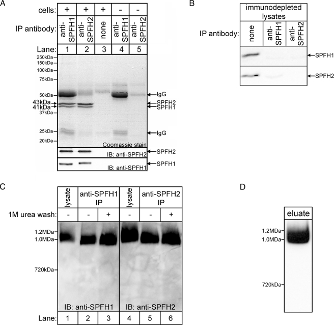
FIGURE 3.
SPFH1 and SPFH2 exist as a complex. A, anti-SPFH1 (lane 1) or anti-SPFH2 (lane 2) IPs fromαT3-1 cell lysates prepared in 1% Triton X-100-containing lysis buffer were subjected to SDS-PAGE and then Coomassie Blue stained (upper panel) or probed for SPFH2 and SPFH1 (lower panels). Controls with either no antibody (lane 3) or no cell lysate (lanes 4 and 5) were also analyzed. B, αT3-1 cell lysates were incubated without or with anti-SPFH1 or anti-SPFH2, and the immunodepleted lysates were subjected to SDS-PAGE and probed for SPFH1 and SPFH2. C, αT3-1 cell lysates (lanes 1 and 4) and immunopurified (IP) SPFH1/2 complex (lanes 2–3 and 5–6) were subjected to BN-PAGE and probed for SPFH1 or SPFH2. The IPs were either not washed (lanes 2 and 5) or were washed (lanes 3 and 6) with 1 m urea prior to elution of SPFH1/2 complexes. D, αT3-1 cells were treated with 100 nm GnRH for 3 min, anti-IP3R1 IPs were prepared, co-purifying SPFH1 and SPFH2 were allowed to dissociate from the IPs, and the eluate was subjected to BN-PAGE and probed for SPFH2. IB, immunoblot.

