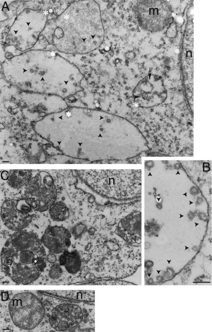
Figure 7.
PIKfyveK1831E expression induces formation of enlarged MVB-like structures with reduced number of internal vesicles and membrane whorls. Doxycycline-treated PIKfyveK1831E-expressing (A and B), PIKfyveWT-expressing (C), or parental HEK293 cell lines (D) were observed by electron microscopy as described under MATERIALS AND METHODS. Enlarged MVB-like compartments (asterisks) with relatively fewer lumenal vesicles (arrowheads) and membrane whorls (arrows) are seen in PIKfyveK1831E-transfected cells (A and B). Depicted are the lumenal vesicles relatively to MVB limiting membrane at higher magnification (B). Micrographs are representative of >100 cells observed in two independent experiments. n, nucleus; m, mitochondria. Bar, 0.1 μm.

