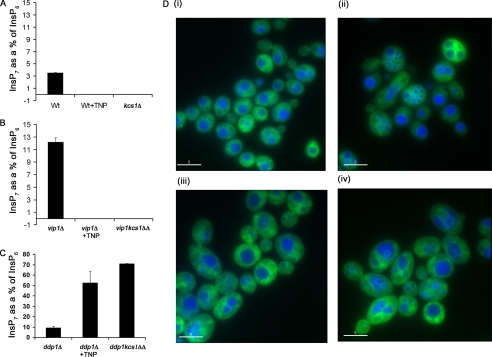
FIGURE 9.
Inhibition of InsP7 formation in WT yeast cells mimics the defectin vacuole morphology reported for ipk2ΔIPK2 and kcs1Δ strains. WT, kcs1Δ, vip1Δ, and kcs1vip1ΔΔ cells were grown in yeast minimal medium supplemented with 50 μCi/ml [3H]inositol overnight at 30 °C. Inositol phosphates were extracted using a protocol similar to that with mammalian cells. HPLC traces are shown in Fig. S3. For vacuole labeling experiments, WT, ipk2Δ, or kcs1Δ strains of haploid yeast cells were grown in YPD. WT cells were grown in the presence of vehicle (DMSO) or TNP. They were then stained with cell tracker CMAC (blue), which stains the vacuolar lumen and membrane marker MDY-64 (green) according to the manufacturer's instructions. Shown is a comparison of InsP7 levels in WT, WT plus TNP, and kcs1Δ cells (A); vip1Δ, vip1Δ+ TNP, and kcs1vip1ΔΔ cells (B); and ddp1Δ, ddp1Δ+TNP and ddp1kcs1ΔΔ cells (C). D (i), WT cells grown in the presence of vehicle (DMSO). D (ii), WT cells grown in the presence of TNP (10 μm). D (iii), ipk2Δ grown in the presence of vehicle (DMSO). D (iv), kcs1Δ grown in presence of vehicle (DMSO).

