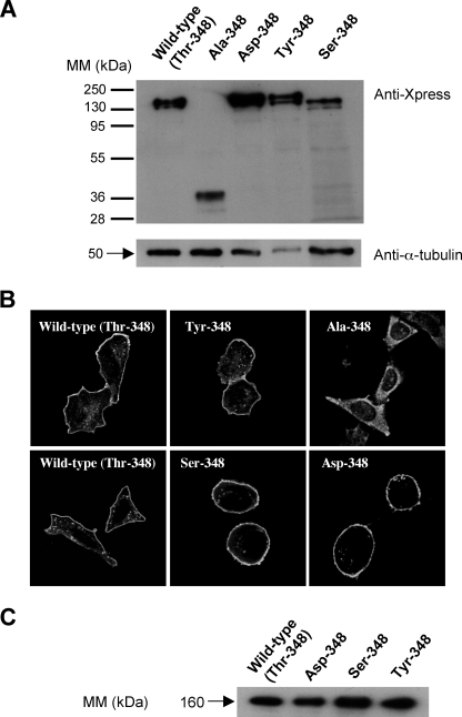
FIGURE 3.
Expression of histidine-tagged wild type and mutated mouse recombinant APAs. A, CHO cells transitory transfected with wild type or mutated His-APA constructs were analyzed by SDS-PAGE 8% and Western blotting with a mouse anti-X-press antibody. The membrane was then stripped and reprobed with a mouse anti-α-tubulin. B, transfected CHO cells stably expressing wild type and mutated His-APA constructs were fixed and immunolabeled with a rabbit polyclonal anti-(rat-APA) serum, which was detected with a cyanin 3-conjugated anti-rabbit secondary antibody. Immunofluorescence was observed by confocal microscopy. The bar indicates 20 μm. C, crude membrane preparations were solubilized and subjected to metal affinity chromatography, as previously described (18). Material eluted (0.25 μg of protein) from the column was analyzed by SDS-PAGE 7.5% and Western blotting with a mouse anti-X-press antibody. Immune complexes were detected by enhanced chemiluminescence. MM, molecular mass.

