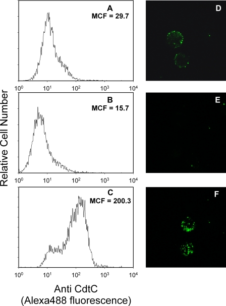
FIGURE 1.
Cdt holotoxin association with Jurkat cells is dependent upon the presence of cholesterol. Jurkat cells were exposed to medium (panels A and D) or 5 mm MβCD (panels B and E (Sigma)) for 30 min. Cells were washed, and some of the MβCD-treated cells were incubated with cholesterol-saturated MβCD (0.5 mm) for 30 min (panels C and F). Cells were incubated with Cdt holotoxin (2 μg/ml) for 1 h, washed, and treated with control murine IgG (data not shown) or anti-CdtC monoclonal antibody conjugated to Alexafluor 488. Jurkat cells were then analyzed by flow cytometry (panels A–C), or images of cells were taken on a Bio-Rad Radiance 2100 Confocal Microscope (Bio-Rad); representative images are shown in panels D–F. Results are representative of three experiments. The relative level of cholesterol in 107 Jurkat cells was compared by semiquantitative TLC. Cholesterol was identified based on calculated Rf values and color after detection by charring with sulfuric acid/EtOH (1:1 vol:vol). The intensity of cholesterol for each sample was compared with the intensity of known standards using digital densitometry; values were 11.8, 0.37, and 20.7 μg cholesterol/107 cells for control, MβCD-treated, and cholesterol-saturated MβCD-treated cells, respectively.

