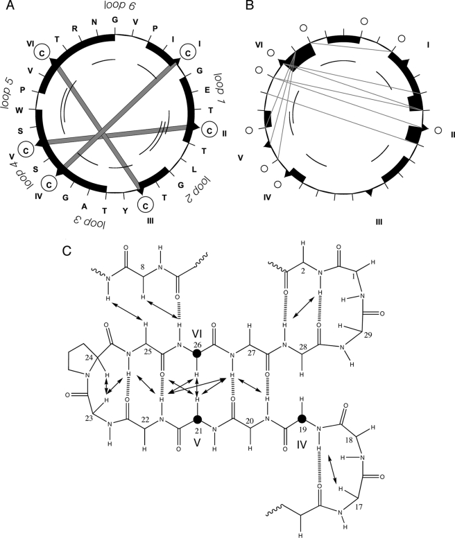
FIGURE 2.
Summary of structurally informative NMR data for varv F. Panel A shows the sequence (in a clockwise direction) and disulfide bonds as thick shaded lines between the cysteine residues (circled). Panels A and B, respectively, show short-range dαN NOEs and dNN NOEs. Black boxes represent sequential NOEs (the size of the box relates to the size of the NOE), whereas arcs represent NOEs between residues separated by 2 or 3 residues in the sequence. In panel B long-range NOE connectivities are shown as gray lines. Panel C focuses on the secondary structure. To illustrate the position of the residues in the cystine knot, three cysteines (CysIV, CysV, and CysVI) are highlighted with bold circles. The NOEs and predicted hydrogen bonds are included.

