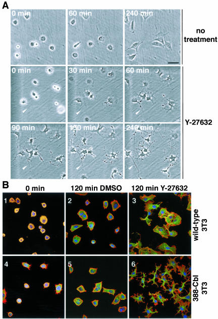
Figure 4.
Inhibition of ROCK induces polarity and microtubule-rich extensions in freshly plated 388-Cbl–expressing NIH 3T3 fibroblasts. (A) Phase contrast microscopy images of freshly plated 388-Cbl–expressing cells were collected during incubation in the absence (1–3) or presence of Y-27632 (4–9). The position of a cytoplasmic extension has been indicated by the arrowhead. Bar, 50 μm. (B) Confocal immunofluorescence microscopy images of TRITC-phalloidin (red), anti-tubulin antibody (green), and Hoescht (blue) staining of wild-type (1–3) and 388-Cbl–expressing NIH 3T3 cells (4– 6) were collected 30 min after replating (1 and 4), or after an additional 120 min of incubation in the absence (2 and 5) or presence of Y-27632 (3 and 6). Bar, 50 μm.

