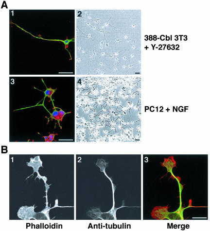
Figure 5.
Inhibition of ROCK induces polarity and extensions in freshly plated 388-Cbl–expressing NIH 3T3 fibroblasts. (A) Confocal immunofluorescence microscopy images of TRITC-phalloidin (red), anti-tubulin antibody (green), and Hoescht (blue) staining (1 and 3), and phase contrast images (2 and 4) were collected 20 h after replating of 388-Cbl–expressing NIH 3T3 cells in the presence of Y-27632 (1 and 2), or 3 d after plating of PC12 cells in the presence of NGF (3 and 4). Bar, 50 μm. (B) Confocal immunofluorescence microscopy images of TRITC-phalloidin (1), anti-tubulin antibody (2) of 388-Cbl–expressing NIH 3T3 cells were collected 6 h after replating in the presence of Y-27632. A merged image of the TRITC-phalloidin and anti-tubulin signals is shown in 3. Bar, 12.5 μm.

