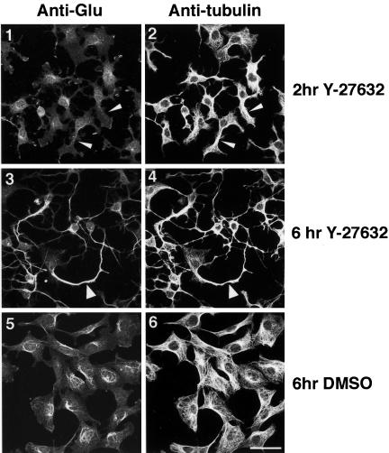
Figure 8.
Microtubule bundles formed by ROCK inhibition are enriched in detyrosinated tubulin. Confocal immunofluorescence microscopy images of anti-Glu-tubulin (1, 3, and 5) and anti-β-tubulin (2, 4, and 6) staining of 388-Cbl–expressing cells were collected after 2 h (1 and 2) and after 6 h (3–6) of replating in the presence (3 and 4) and absence (5 and 6) of Y-27632. Non-Glu– and Glu-tubulin–containing microtubule bundles are indicated by narrow and wide arrowheads, respectively. Bar, 50 μm.

