FIGURE 2.
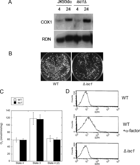
Lack of intrinsic mitochondrial defects in Δisc1. A, mitochondrial DNA content of WT and Δisc1. The genomic DNA of WT and Δisc1 was obtained from 4 and 24 h after inoculation and analyzed by Southern blotting with the mitochondrial COX1 and nuclear RDN1 probe. B, WT and Δisc1 were incubated in medium containing ethidium bromide for 8 h and plated onto YPD plates. The plates were incubated at 30 °C for 2 days. Plating onto YPGE plates did not produce any colonies (data not shown). C, oxygen consumption rate of the isolated mitochondria measured as described under “Experimental Procedures.” The results are represented as the mean ± S.E. of at least two independent experiments. D, reactive oxygen species of WT cells with or without treatment with α-factor (final 11.9 μm at 30 °C for 1.5 h) and Δisc1 cells. The cells were mixed with 40 μm 2′,7′-dichlorofluorescein diacetate and applied to FACS analysis.

