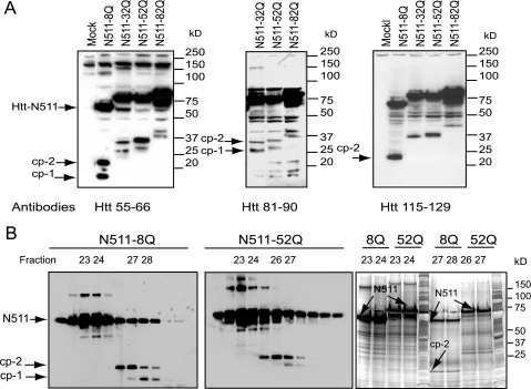
FIGURE 2.
Expression and purification of Htt fragments in HEK293 cells. A, epitope mapping of the cp-1/cp-2 fragments. Western blot of total cell extracts (30 μg/lane) from HEK293 cells transfected with either N511-Htt constructs with different poly(Q) lengths, or cells transfected with empty vector (Mock). Htt-N511 and cp-1/cp-2 fragments (arrows) were detected with an antibody to residues 55-66 of Htt (left panel), with antibody to residues 81-90 of Htt (middle panel), or with antibody to residues 115-129 of Htt (right panel). HEK293 cells transfected with expanded Htt constructs produce fragments similar to the cp-1/cp-2 fragments observed previously in PC12 cells (30). B, purification of the cp-1/cp-2 fragments for mass spectrometry. Western blotting analysis of 1/10 aliquots of fractions from size chromatography of FLAG immunoprecipitates of either Htt-N511-8Q (left panel), or Htt-N511-52Q (middle panel) are expressed in HEK293 cells. Htt proteins, marked by arrows, were detected with antibody to FLAG. Indicated fractions containing either the cp-1/cp-2 fragments or in Htt-N511, were separated on SDS-PAGE, and stained with silver protein stain (right panel). Htt bands (indicated with arrows) corresponding to cp-1/cp-2 fragments, and bands containing only Htt-N511 were excised from the gel for mass spectrometry.

