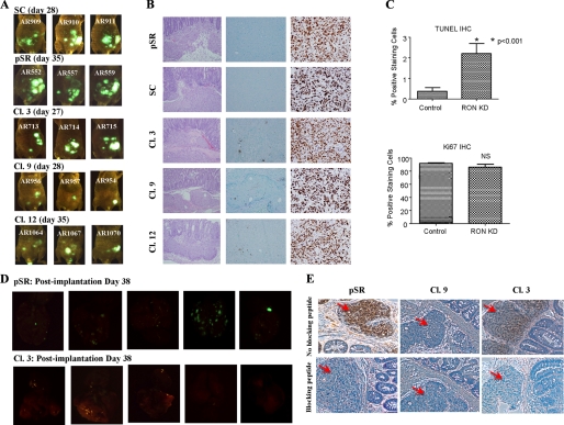
FIGURE 7.
Exponentially growing cells (5 × 10⁶) were inoculated subcutaneously in athymic nude mice. The xenografts formed by the control cells and Ron knockdown clones were used for subsequent orthotopic implantation. A, fluorescence imaging of the animals on the indicated days. B, primary tumors established from the control and RON knockdown cells were processed for hematoxylin and eosin (left panel), TUNEL (middle panel), and Ki67 (right panel) staining. Both TUNEL and Ki67 images were captured at ×20 magnification. C, primary tumors established from the control and RON knockdown cells were analyzed to assess the proliferation and apoptotic rate. NIH Image J software was utilized to enumerate positive staining cells and total number of cells. The percentage of positive staining cells was calculated. Error bars indicate S.E. D, 38 days post-implantation, animals were euthanized. Organs were explanted and imaged. E, immunohistochemistry was performed for pAKT (S473) on primary tumors of control or Ron knockdown cells. The blocking peptide was used to show the specificity. The red arrows indicate tumor cells.

