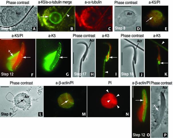
Figure 3.
Development of the acroplaxome during rat spermiogenesis by using antibodies to K5 and β-actin. (A–C) A step 8 spermatid displays a top view of the acrosome (acr, A). The 1-μm-wide marginal ring of the acroplaxome (arrow) and an overlapping spermatid tail (arrowhead) are K5 immunoreactive (B). α-Tubulin–stained microtubules of the manchette are seen eccentric to the acroplaxome in C. (D and E) Step 8 spermatid subjected to hypotonic treatment to remove the cytoplasm and stained with anti-K5 serum and propidium iodide (PI). The K5-immunoreacted acroplaxome (arrow) remains attached to the spermatid nucleus. (F and G) Step 12 spermatid displaying the K5 immunoreactive manchette (crossed arrow) and acroplaxome (arrow) attached to the elongating PI-stained spermatid nucleus. (H and I) Step 17 spermatid. The K5 stained component of the acroplaxome occupies a caudal site (arrow), and the spermatid tail (arrowhead) displays similar immunoreactivity. (J and K) Step 18 spermatid showing a more caudal localization of K5 immunoreactivity. An immunoreactive spermatid tail (arrowhead) is seen in the field. (L–N) Step 8 spermatid is devoid of cytoplasm after hypotonic treatment but retains the acrosome (acr, arrow). The marginal ring of the acroplaxome (arrow) is β-actin immunoreactive (compare with E). (N) Strong and uniform cap-like PI nuclear staining at the acroplaxome site (denoted by arrowheads). (O and P) Step 12 spermatid. The arrow points to the β-actin immunoreacted acroplaxome. The manchette (crossed arrow in P) seems essentially nonimmunoreactive by immunofluorescence (compared with F and G). Bars, 5 μm.

