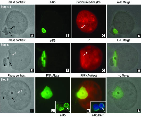
Figure 4.
The attachment of the rat spermatid acroplaxome to the nuclear envelope is not disrupted by Triton X-100 extraction before fixation. (A–D) A step 4 to 5 spermatid displays in A its acrosome (arrow) lodged in a shallow depression of the nucleus (crossed arrow). The Golgi apparatus (arrowhead) remains attached to the acrosome. The K5-immunoreactive acroplaxome is shown in B. (C) A linear profile of propidium iodide-stained nuclear material is seen along the shallow depression (crossed arrow). (E–H) Step 6 spermatid displays the same features observed in the A–D sequence except that the linear propidium iodide stained nuclear material is more extensive and matching the length of the acroplaxome. (I–L) Step 6 spermatid showing a frontal view of the acrosome (arrow) surrounded by a dense edge (crossed arrow) and associated Golgi apparatus (arrowhead). (J) PNA-Alexa Fluor 488 conjugate uniformly stains the acrosome and the proximal components of the Golgi apparatus. In contrast, the insets in panels J and K show the marginal ring of the K5-stained acroplaxome (inset in J) overlapping the 4,6-diamidino-2-phenylindole-stained nuclei (inset in K) not visualized after PNA-Alexa Fluor 488 staining. Bars, 3 μm.

