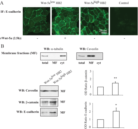
FIGURE 2.
A, immunofluorescence (IF) studies of E-cadherin membrane localization in human breast epithelial cells (HB2) with varying Wnt-5a protein expression levels or in HB2 Wnt-5alow cells treated with recombinant Wnt-5a (rWnt-5a)(n = 3). B, membrane fractionation experiments. The purity of the membrane fractions (total cell lysate (total), membrane fraction (MF), and cytosol (cyt)) was analyzed with α-tubulin and caveolin antibodies (upper panel). Western blot analysis of E-cadherin and β-catenin protein expression levels in membrane fractions from human breast epithelial cells with varying Wnt-5a protein expression levels (lower panels). To ensure equal loading, protein measurements were done using a Coomassie® protein assay reagent (Pierce). The blots shown are representative of several separate experiments and OD measurements of the band intensities were performed to quantify the differences (right panel, histograms). Error bars indicate S.E., n = 16; *, p < 0.05; **, p < 0.01. WB, Western blot.

