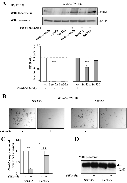
FIGURE 7.
A, co-immunoprecipitation of E-cadherin with different forms of FLAG-tagged β-catenin (35). HB2 Wnt-5alow cells were transfected with FLAG-tagged wild type (wt) β-catenin, Ser-45Δ-mutated β-catenin, and Ser-33Δ-mutated β-catenin. Immunoprecipitation was performed using anti-FLAG antibodies. Due to a decreased degradation of β-catenin harboring the Ser-45Δ and Ser-33Δ mutations, FLAG β-catenin had to be set to equal levels in the blots by a pre-Western blot (WB) analysis after which the level of coprecipitated E-cadherin compared with FLAG β-catenin in the final blot could be measured using OD measurements. The histogram thus shows the relative levels (OD E-cadherin/OD FLAG-β-catenin) of bound E-cadherin to FLAG-tagged wild type β-catenin as compared with FLAG-tagged Ser-45Δ and Ser-33Δ β-catenin, under normal (left) or Wnt-5a stimulating (right) conditions (***, p < 0.001 compared with wild type, S.E., n = 7). B, Matrigel-invasion assay. HB2 Wnt-5alow cells were transfected with FLAG-tagged Ser-33Δ β-catenin or Ser-45Δ β-catenin. After a 48-h transfection period the cells were detached, counted, and 25,000 cells/chamber were allowed to invade Matrigel invasion chambers in the absence or presence of rWnt-5a in the upper chamber. C, the histogram (representing B) shows the fold-reduction in number of invaded cells upon rWnt-5a treatment for both Ser-33Δ β-catenin- and Ser-45Δ β-catenin-transfected cells (**, p < 0.01; *, p < 0.05; S.E.; n = 3). D, the levels of FLAG-tagged β-catenin protein were similar between the samples in B and C.

