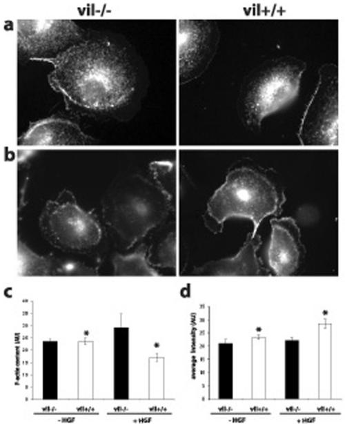
Figure 5.
Villin-expressing cells present a decrease of the F-actin content and a higher level of barbed ends at the leading edge of MDCK cells upon HGF stimulation. Free barbed ends were visualized by rhodamine G-actin incorporation before (a) and after 6 h of HGF treatment (b). The amount of free barbed ends at the leading edge of the cells was evaluated by quantitation of the average fluorescence intensity using MetaMorph software (d). Eighteen vil-/- and 24 vil+/+ unstimulated cells were analyzed. Twentyseven vil-/- and 30 vil+/+ cells after 6 h of HGF treatment were analyzed (*p = 0.004). The F-actin content was evaluated by measuring the fluorescence intensity of TRITC-palloidin–labeled actin filaments in MDCK cells before and after 3 h of HGF treatment (c). The amount of total protein was the same in vil-/- and vil+/+ cells; n = 3 experiments.

