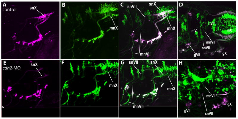
Figure 4. The effects of cdh2 knock-down are specific to the afferent fibers.
A cross between tg(PB4:GVP;UAS:kaede) and tg(Isl1:eGFP) fish show that snVII central processes project aberrantly even though mnVII projects normally in cdh2-MO injected embryos. A-D depict a control embryo, E-H depict a cdh2-MO injected embryo. Note also, neurons in the CSG appear scattered in cdh2-MO injected embryos (E-G). D and H are dorsolateral views showing the positioning of the motor nuclei. In all images, the sensory circuits are labeled with photoconverted kaede, shown as magenta, and all motor circuits (and some sensory neurons) contain GFP labeling that is shown in green. Cells expressing both proteins are seen as white. Images are from 96hpf embryos. Rostral is left, dorsal is top.

