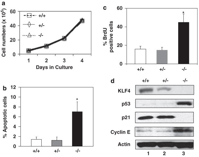
Figure 1.
Growth characteristics of Klf4+/+, Klf4+/− and Klf4−/− mouse embryonic fibroblasts (MEFs) in culture. (a) Cells were plated at 105 cells per 60-mm plate. Three plates were counted at each time point and the values represent the mean number of cells per dish. N = 3. (b) The percentages of apoptotic cells 1 day after seeding were measured from the sub-G1 population of cells during flow cytometry. N = 3; *P<0.05 compared to Klf4+/+ MEFs. (c) DNA synthesis was measured by the incorporation of bromodeoxyuridine (BrdU) into replicating cells. Shown are the percentages of cells that stained positive for BrdU. N = 3; *P<0.05 compared to Klf4+/+ MEFs. (d) Western blot analysis of KLF4, p53, p21, cyclin E and β-actin of proteins isolated from cells at 1 day after seeding. Shown are the representative results of four separate experiments.

