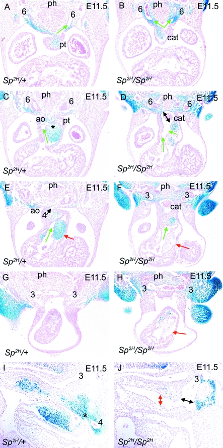
Fig. 3.
NCC distribution in Sp2H embryos at E11.5, as outflow tract septation is initiated. In each case, the direction of blood flow through the outflow tract is denoted by green arrows. (A,C,E,G) Transverse sections show that fusion between the dorsal wall of the aortic sac and the distal outflow tract cushions is underway (* in C) in Sp2H/+ embryos, both of which are filled with βgal-expressing NCC. As a consequence, an aortic (green arrow in C) and pulmonary (green arrow in E) channel are apparent in the outflow tract by this stage. The 3rd, 4th and 6th arch arteries are all apparent, although the right 4th and 6th arteries are regressing by this stage. (B,D,F,H) Fusion between the dorsal wall of the aortic sac and the distal outflow cushions does not occur in the Sp2H/Sp2H embryo (double-headed arrow in D). A reduction in the mesenchyme between the pharynx and the aortic sac is still marked in Sp2H/Sp2H embryos. The outflow cushions are deficient in NCC and the proximal regions appear acellular (red arrows in F,H). Despite this, two channels are apparent in the Sp2H/Sp2H outflow tract (green arrows in D). In this embryo, left and right 3rd and 6th arch arteries can be seen, but neither right nor left 4th arch arteries. (I,J) Sagittal sections show the fusion point (asterisk) between the dorsal wall of the aortic sac and the outflow cushions in Sp2H/+ embryos (I). In Sp2H/Sp2H embryos, the outflow tract cushions are hypoplastic with a marked reduction in βgal-stained cells and there is no fusion with the dorsal wall of the aortic sac (black double-headed arrow in J). The outflow cushions are widely separated (red double-headed arrow in J) and the 3rd arch artery appears dilated. ao, aorta; cat, common arterial trunk; ph, pharynx; pt, pulmonary trunk.

