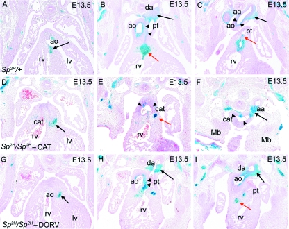
Fig. 4.
NCC distribution in Sp2H fetuses at E13.5, after outflow tract septation. (A–C) NCC are found at the fusion point between the outflow cushions and the ventricular septum (arrow in A), the proximal outlet septum (red arrow in B), the great arteries including the walls of the ductus arteriosus (black arrow in B), the aortic arch (black arrow in C) and the separating walls of the aorta and pulmonary trunk (arrowheads in B). βgal-expressing NCC are also found in the pulmonary valves (red arrow in C). (D–F) In Sp2H/Sp2H fetuses with common arterial trunk, NCC are found in the proximal outflow region (arrow in D). Although there are NCC in the aortic arch (arrow in F), there are fewer βgal-expressing NCC in the proximal walls of the common trunk (arrowheads in E and F). A focus of βgal-expressing cells is found in the region where the proximal outlet septum would normally be found (red arrow in E). The aortic arch (F) is seen at the same axial level as the developing mandible, showing its cervical position. (G–I) In Sp2H/Sp2H fetuses with double outlet right ventricle, NCC are found in the proximal outflow region (black arrow in G) and the walls of the great arteries including the ductus arteriosus (black arrow in H), the aortic arch (black arrow in I) and the separating walls of the aorta and pulmonary trunk (arrowheads in H). Although the proximal outlet septum is hypoplastic, a small focus of NCC can be seen (red arrow in I). aa, aortic arch; ao, ascending aorta; cat, common arterial trunk; da, ductus arteriosus; lv, left ventricle; mb, mandible; pt, pulmonary trunk; rv, right ventricle.

