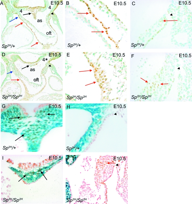
Fig. 5.
Isl1 immunostaining of SHF cells and βgal expression by NCC in Sp2H embryos at E10.5. (A–C) The dorsal wall of the aortic sac between the 4th and 6th arch arteries, which will fuse with the distal outflow cushions, is filled with Isl1-expressing cells in Sp2H/+ embryos (black arrow in A). Isl1-immunostained cells are also found in the outer layer of the lateral sides of the aortic sac (blue arrow in A) and in the outflow myocardium (red arrows in A,B), in each case forming a single cell layer. (D–F) In Sp2H/Sp2H embryos, few Isl1-expressing cells can be seen in the dorsal wall of the aortic sac (black arrow in D). Isl1 immunostaining is seen in the lateral sides of the aortic sac (blue arrows in D) and the outflow myocardium (red arrows in D,E). However, the staining in the myocardium reveals that the cells are multilayered (red arrows in E), rather than forming a monolayer as in the Sp2H/+ embryos (red arrows in B). In addition, ectopic Isl1-expressing cells are found in the thickened walls of the aortic sac (red arrows in F). (G,I) NCC and SHF cells are both found in the dorsal wall of the aortic sac, between the origins of the 4th and 6th arch arteries in Sp2H/+ and Sp2H/Sp2H embryos (black arrows), although the numbers of each are much reduced in the hypoplastic structure of the Sp2H/Sp2H embryo. (H,J) Whereas blue-stained NCC are found in the mesenchyme of the aortic sac and distal outflow cushions in Sp2H/+ embryos, they are absent from these latter areas in Sp2H/Sp2H embryos. Ectopic Isl1-expressing SHF cells can be seen in the thickened lateral walls of the aortic sac in Sp2H/Sp2H embryos (red arrow in J). (A,C,D,F,H,J) The black arrowhead marks the pericardial reflections and thus is a landmark which allows comparison between different sections and embryos. as, aortic sac; oft, outflow tract.

