Fig. 4.
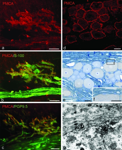
Immunolocalization of PMCA-ATPase in the periodontal ligament (a–c) and trigeminal ganglion (d–g). (a,b) Double labeling with PMCA and S-100 protein shows that the axon terminals have both PMCA and S-100 immunoreactions; however, the cell bodies do not express any co-localization. (c) PGP9.5 positive axonal processes are detected with PMCA immunoreaction. (d–f) In the trigeminal ganglion, the PMCA-positive reactions exist along the outlines of neuron cells. These reactions are found in the cell membrane of the neuronal cell. (g) Immunoelectron micrograph of the trigeminal ganglion. Electron-dense immunopositive products gather on the cell membrane, especially in a finger-like projection. Scale bars: 25 µm (a–e), 20 µm (f), 0.15 µm (g).

