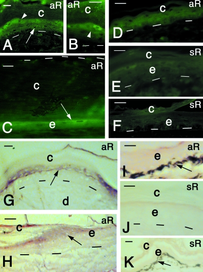
Fig. 7.
In situ hybridization results on turtle skin after immunofluorescence (A–F) and alkaline phosphatase-colorimetric-based methods (G–K). (A) Reactive cells in the upper suprabasal (arrow) and pre-corneous (arrowhead) layers. Lateral area of carapace scute. Bar, 15 µm. (B) Close-up on reactive spindle-shaped cells (arrowhead) of the pre-corneous layer. Lateral areas of carapace scute. Bar, 20 µm. (C) Central area of carapace scute showing the thick corneous layer (delimited superiorly by dashes) and the thin, reactive cell of the epidermis (arrow). Bar, 10 µm. (D) Relatively soft epidermis of the limb showing some weak reactivity in the epidermis. Bar, 10 µm. (E) Sense control in the epidermis of a carapace scute. Bar, 15 µm. F, sense control of soft epidermis of limb. Bar, 15 m. (G) Reactive cells (arrow) of the precorneous layers (arrow) in carapace scute. Bar, 10 µm.(H) Reactive cells (arrow) near the hinge region (growing center) of carapace scute. Bar, 20 µm. (I) Non-reactive soft epidermis of the limb. Bar, 15 µm. (J) Sense control of carapace scute epidermis. No reactivity is present. Bar, 10 µm. (K) Sense control of soft epidermis of the limb. No reactivity is present. Bar, 10 m. aR, antisense RNA probe; c, corneous layer; d, dermis; e, epidermis; sR, sense RNA probe. Dashes underlie the basal layer of the epidermis.

