Fig. 5.
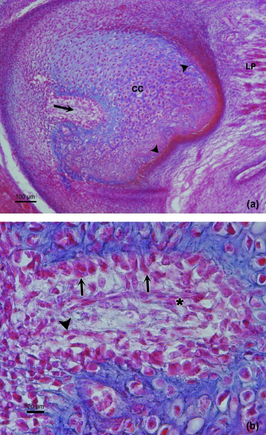
(a) Human fetus Be-503 (48-mm CRL; 10 weeks of development). Azocarmine staining. Frontal section. The posterolateral canal (arrow) deepens into the condylar cartilage (CC). Arrowhead, intramembranous ossification; LP, lateral pterygoid muscle. Bar 100 µm. (b) Human fetus Be-503 (48-mm CRL; 10 weeks of development) Frontal section. Azocarmine staining. The periphery of the posterolateral canal shows cuboid cells in band formation (arrows), associated with the cartilaginous intercellular matrix. In the lower part of the canal the band arrangement is not clearly discernible because of the obliqueness of the section. Inside the canal, mesenchymal cells (arrowhead) and vessels (asterisk) appear. Bar: 20 µm.

