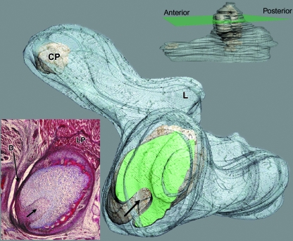
Fig. 8.
3D reconstruction of human fetus JR-6 (80-mm CRL; 12 weeks of development). The upper-right image indicates the section level (green plane). The reconstruction shows the arrangement of the condylar cartilage at the section level labelled in green (arrow, posterolateral vascular canal). The lower-left image shows the histological section that matches the section level pointed out in the reconstruction. L, lingula; CP, cartilage of the coronoid process; LP, lateral pterygoid muscle; D, articular disc. Bar: 200 µm.

