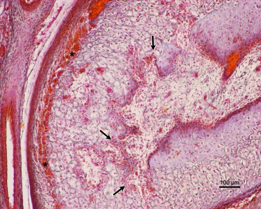
Fig. 10.
Human fetus HL-30 (74-mm CRL; 12 weeks of development). Transverse section. Azocarmine staining. In the lower part of the condylar cartilage there are signs of vascular invasion and cartilage destruction (arrows). Some chondroclasts are visible in the medial part of the condylar cartilage (asterisk). D, articular disc. Bar: 100 µm.

