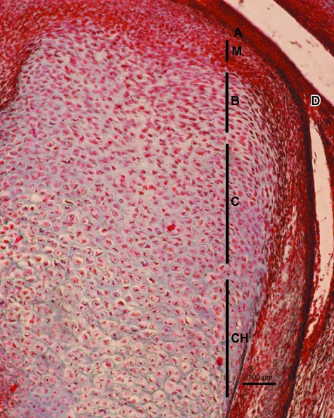
Fig. 11.
Human fetus Be-516 (82-mm CRL; 13 weeks of development). Sagittal section. Haematoxylin-eosin staining. The different layers of the condylar cartilage are visible. A, articular layer; M, mesenchymal layer; B, chondroblastic layer; C, chondrocyte layer, CH, hypertrophic chondrocyte layer. Bar: 100 µm.

