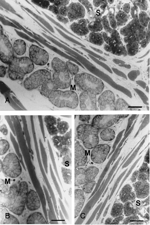
Fig. 1.
Light microscopy showed no significant morphological changes in posterior deep and superficial lingual glands after hypoglossal denervation. (A) Normal serous and mucous glands beneath the circumvallate papilla between striated muscle fibers on the control side. No significant morphological alteration in the glands 3 (B) or 7 days (C) after hypoglossal denervation. S, serous glands; M, mucous glands. Toluidine blue. Bars = 150 µm.

